

1. Introduction
2. Experimental Methods
2.1 Materials
2.2 Shock tube experiments
3. Results and discussions
4. Conclusions
1. Introduction
Materials that can withstand shock waves are crucial for aerospace, defense, energy, and materials science research fields. These materials maintain their integrity and function even under extreme conditions [1,2,3]. The materials undergo phase transitions, fractures, and changes in atomic and molecular arrangements of shock waves which lead to amorphous to crystalline nature and vice versa. Stable materials ensure safety by preventing catastrophic failures in critical applications like spacecraft, aircraft, and military armor. Similarly, materials must remain intact in high-speed aerospace to avoid compromising the craft’s integrity. In energy industries, materials must withstand shock waves from drilling, explosions, or stress without degrading, as failure could lead to hazards. Stable materials also play a key role in research, especially in synthesizing novel compounds or nanomaterials under shock wave studies, where controlling material behavior under high pressures and pressure is essential.
Furthermore, energy storage and catalytic applications under shock waves to enhance material properties—without causing instability—can improve efficiency, performance, and durability [4,5]. The importance of shock wave-resistant materials derives from their ability to maintain reliability, extend lifespan, and assure the safe and efficient operation of technologies in extreme conditions. These materials are critical for maintaining the integrity and performance of various technologies and systems in industries, where they are exposed to high-impact forces, intense pressures, and other extreme conditions such as shock wave propagation on materials [6,7,8].
Consequently, shock wave research has emerged as an innovative field, and gained widespread acceptance due to its distinctive influence and the abundance of publications that provide profound insights into the specific domain of nanomaterials. A wide range of nanomaterials have been the subject of in-depth research in recent decades because of their distinct optical, chemical, electrical, and physical characteristics when subjected to shock waves. Shock wave-loaded cerium oxide nanoparticles exhibit remarkable stability in their crystallographic structure and electronic phase, with no phase transitions detected through X-ray diffraction and Raman spectroscopy analyses. Furthermore, the valence state of Ce4+ remains unchanged according to ultraviolet-visible diffuse reflectance spectroscopy findings. Field emission scanning electron microscopy also confirms the stability of the sample, suggesting potential applications for CeO2 nanoparticles in medical glassware, spacecraft window materials, and protective coatings [9]. Using pressure-driven shock waves at 2.683 MPa, TiO2 nanoparticles changed the structure from the anatase phase to the rutile phase [10]. Shock waves at 2.0 MPa induce a phase transition in Co3O4 nanoparticles to CoO after 150 pulses, confirmed by XRD and Raman analyses. Optical band gap, morphology, and magnetic properties show significant changes, including switchable superparamagnetic behavior due to dynamic recrystallization [11]. Further, the dynamic shock wave experiments on BaCO3 nanoparticles reveal exceptional structural stability, retaining the original Pmcn crystal structure even after 200 shock pulses at Mach 2.2. Analytical results indicate BaCO3 superior shock resistance compared to materials like TiO2 and Co3O4, highlighting its potential for space electronics, optical glasses, condensers, and aerospace pigments [12]. Also, WS2 nanotubes exhibit remarkable resilience, withstanding shear stress up to 21 GPa, despite some defective tips becoming damaged and small WS2 layers extruding. In comparison to carbon nanotubes, WS2 nanotubes demonstrate superior structural stability, suggesting their suitability as durable lubricants under extreme loading conditions [13]. Shock-loaded NiF2 samples show improved crystallite and particle size, a stable rutile phase, and improved electrochemical properties. These improvements make NiF2 ideal for use in supercapacitors in extreme conditions [14].
Here, the semiconducting transition materials, such as ZnO, TiO2, NiO, CuO, Cu2O, VO2, WS2, MoS2, SnO2, and ZrO2, are at the forefront of academic research, significantly influencing the development of data transmitters, frequency converters, and other electronic devices due to their exceptional physicochemical properties [15]. Among these materials, ZnO is a promising semiconductor with a wide bandgap of 3.37 eV and a substantial exciton binding energy of 60 meV, making it highly attractive for various technological applications [16]. The versatility of ZnO nanostructures has been extensively explored, with researchers synthesizing diverse morphologies, including nanorods, nano-leaves, nanodots, and nanoflowers, each with unique properties and applications [17]. ZnO nanostructures are ideal for extreme-condition applications such as optoelectronics, solar cells, sensors, and light-emitting diodes, as they provide radiation resistance, excellent thermal stability, and oxidation resistance [18]. With the growing interest in aerospace applications, the demand for materials that can withstand extreme conditions is increasing. Consequently, ZnO nanostructures are expected to be crucial in space engineering technology. Therefore, it is essential to thoroughly investigate and understand the sustainability and durability of ZnO nanostructures under the extreme conditions encountered in space applications, such as extreme temperatures, radiation, and high-stress conditions.
Many investigations have shown that exposure to high-energy radiation and shock waves significantly alters the optical properties and structure of the materials. For instance, Hammam et al. reported that the optical bandgap energy of ZnO increases with elevated gamma radiation, which they attributed to variations in particle size. Similarly, the optical transmittance and bandgap of ZnO are also significantly influenced by laser irradiation [19]. Furthermore, a redshift in the UV emission band of ZnO was subjected to third harmonic laser pumping, a phenomenon they ascribed to the occurrence of exciton recombination and electron-hole plasma recombination [20]. Shock wave recovery experiments on ZnO nanorods reveal their high resilience in visible light and negligible transmittance decline in ultraviolet under shock conditions. Additionally, optical absorption exhibits only a minor increase with shock pulses, while Raman spectroscopy verifies the stable crystalline structure, suggesting the suitability of ZnO nanorods for space applications [21]. The researcher’s investigations on the shock wave recovery of ZnO nanorods demonstrated remarkable stability in the material’s molecular, optical, structural, and morphological properties under 150 pulse shock loads. While a slight blue shift in the UV-vis spectrum was observed at 200 pulses, X-ray diffraction analysis revealed no structural changes. These findings indicate that ZnO nanorods are a suitable candidate for military and aerospace applications [22]. Furthermore, ZnO nanorods grown on stainless steel surfaces are extremely durable when exposed to dynamic shock waves, retaining their crystalline structure and shape despite major changes in chemical composition and luminescence properties. According to these results, ZnO nanostructures can withstand pressures lower than 2 MPa and temperatures as high as 8000 K, making them ideal for use in space engineering technologies [23]. This study combined high-dose proton irradiation with rapid thermal annealing to improve the performance of ZnO thin-film transistors. Rapid thermal annealing improved the electrode-channel interface but decreased off-state control because of the decreased resistivity. By increasing resistivity and dramatically enhancing field-effect mobility, proton irradiation solved this problem and produced better electrical properties and overall TFT performance [24]. Also, the author reported that the high enthalpy shock waves on graphitic carbon nanoparticle (GCNP) films lead to material loss and growth of crystalline nanostructures, especially cubic diamond structures, with increased test gas pressure. This treatment enhances film quality and creates hierarchical nanostructures on the surface. These findings support potential applications in space and chemical engineering [25].
ZnO nanomaterials are stable and highly suitable for military and aerospace applications due to their high thermal, chemical, and mechanical stability, enabling them to withstand harsh environments. But the response for repeated exposure to such environment is less explored in the literature. Hence, the present research gives an insight on this problem to show that ZnO is a potential material even for repeated exposure of shock waves to ensure a long-term performance to challenging military and aerospace applications. In the present study to investigate the stability of ZnO nanoparticles under extreme conditions to assess their potential uses in extreme environments, a number of sock pulses such as 100, 200 and 300 are loaded on the sample. X-ray diffraction(XRD) and scanning electron microscopy (SEM) are utilized to investigate the impact of the shock waves on ZnO samples.
2. Experimental Methods
2.1 Materials
Hexagonal-shaped zinc oxide nanoparticles with a 97% purity level were purchased for the shock wave investigation from Sigma Aldrich in South Korea. In addition, the nanopowder was exposed to different shock wave treatments, and the materials’ morphological and structural properties were examined.
2.2 Shock tube experiments
The manually operated tabletop shock tube is a specialized apparatus predominantly used in academic and research settings to generate controlled shock waves with distinct characteristics. Generally, the shock tube consists of three primary components: a driver, a driven section, and a diaphragm. When the gas fills the driver section, the diaphragm ruptures and the resulting shock wave propagates through the driven section. For a clearer understanding, the schematic diagram of the shock tube is presented in Fig. 1.
In the present experiment, open-end wall method is used (i.e the sample is placed outside the shock tube). The shock pulse is a pocket of energy characterized by abrupt increase of pressure and temperature lasting for a very short span of time in the order of microsecond to millisecond. Whenever the diaphragm ruptures, one shock pulse is generated and propagates through the driven section and strikes the sample which is placed 1cm away from the open end of the driver tube. Air is used in both the driver (8 bar) and driven tube (atmosphere pressure) with Mach number of 1.5. Hence, we analyzed the material properties using various shock pulses, such as control, 100, 200, and 300 shock pulses [26].

3. Results and discussions
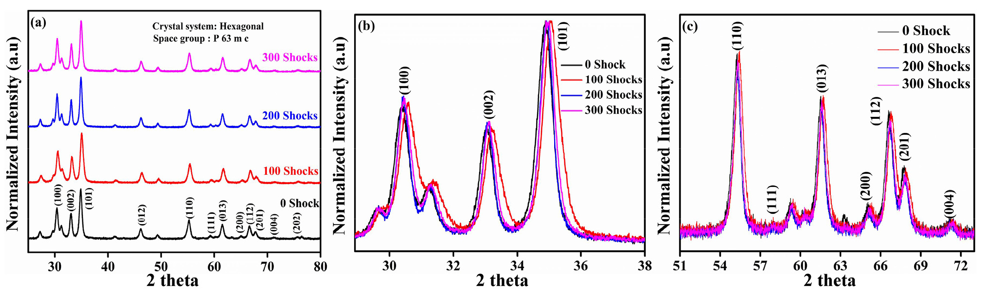
The powder X-ray diffraction pattern depicted in Fig. 2 confirms that the hexagonal wurtzite structure of ZnO nanopowder is preserved for both the control and various shock-loaded samples. X-ray diffraction (XRD) analysis was performed using a multi-purpose X-ray diffractometer (XRD-6000, Shimadzu, Kyoto, Japan) at Intelligent Construction System Core-Support Center, Keimyung University, Republic of Korea. The observed diffraction peaks at 31.88°, 34.56°, 47.68°, 56.76°, 63.06°, 66.51°, 68.14°, 69.30°, 72.90°, and 77.23° correspond to the Miller indices [(00), (002), (102), (110), (103), (200), (112), (201), (004), (202)] from the PDF card number 96-210-7060. These characteristic diffraction peaks indicate that the ZnO nanopowder maintains its crystalline structure even after being subjected to various shock-loading conditions [27] is shown in Fig. 2(a). The prominent peak at the 101 planes suggests that the ZnO nanopowder is predominantly orientated along the 101 directions. As a result, ZnO displays a hexagonal wurtzite structure, which is the most stable phase under shock conditions. Moreover, the shock wave does not trigger any phase transformation. Additionally, no new peaks are detected, and the existing peaks remain unaffected under various shock-loaded conditions. This indicates that the ZnO nanopowder is highly stable and resistant to structural changes even under shock waves [28].
For better understanding, we have chosen a few diffraction peaks and calculated their full width at half maximum (FWHM), analyzing the peak intensity. We have examined the peak intensity for various shock waves applied to different planes of ZnO, as shown in Fig. 2(b), (c). We observed no changes between the control and shock-loaded samples, and the peak intensity remained nearly the same, indicating that the crystallinity of ZnO remained unchanged under shock loading. However, the slight shift in peak position towards higher angles might suggest sudden changes in the fusion and fission of ZnO crystal grain boundaries during shock wave propagation. Additionally, the shift in diffraction peaks shows that a significant density of defects, including stacking faults, vacancies, and dislocations, are introduced into the crystal lattice by ZnO nano powder [21,22].
The analysis of the full width at half maximum of the diffraction peaks indicates that the control samples exhibit a narrower FWHM, while the FWHM increases slightly with increasing shock pulses of 100, 200, and 300 simultaneously is shown in Fig. 3.
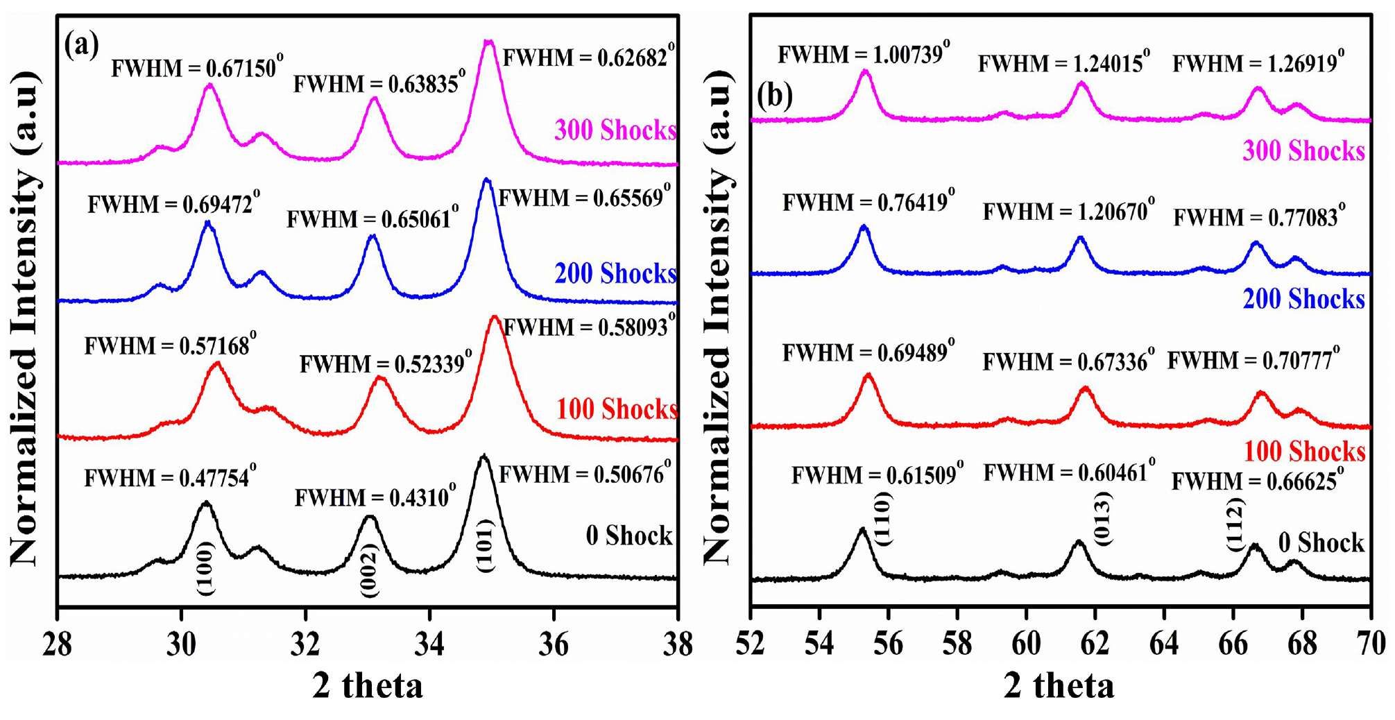
Similarly, the width of all the diffraction peaks demonstrates a slight increase with the increasing shock pulses and this change in peak width leads to peak broadening. The intense mechanical strain generated by shock waves results in distortions in the lattice parameters, which in turn leads to peak broadening and changes in intensity. The degree of intensity changes can reflect the extent of strain, with stronger shocks typically resulting in more pronounced modifications. These observations suggest that the increasing shock pulses has a measurable impact on the microstructural properties of the samples, as evidenced by the changes in the diffraction peaks. The propagation of shock waves through materials induces high pressure and strain within the crystal lattice, leading to grain fragmentation, particularly in nanomaterials. Generally, smaller grains have more grain boundaries, which increases the full width at half maximum due to the reduced crystalline size. Deformation and fragmentation of grains can significantly alter the microstructure and mechanical properties of polycrystalline materials [29].
Fig. 4 represents the calculated lattice parameter and cell volume for the control and shock-loaded samples. calculated using ‘unit cell’ software. The lattice parameter and cell volume decreased for the control and shock-loaded 100 and 200 samples. However, the volume and lattice parameter slightly increased for the 300 shocked samples is shown in Table 1.
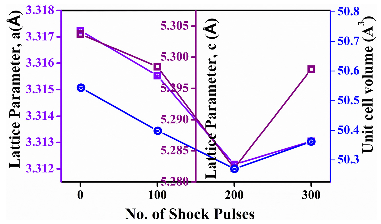
Table 1.
Volume and lattice parameter of ZnO nanomaterials.
Under shock-load conditions, no significant changes were observed. The change in volume and lattice parameters leads to defects like vacancies and interstitials, as well as slight strain in the crystal lattice. This can result in a slight expansion of the lattice parameter and cell volume as atoms are displaced from their equilibrium positions, effectively stretching the lattice. By 300 shocks, the lattice may reach a point where defect accumulation saturates, leading to lattice relaxation or even partial annealing of defects.
The field emission scanning electron microscope was employed to investigate the morphological properties of stable hexagonal zinc oxide nanopowder in both control and shock-loaded samples. The control and shock-loaded samples displayed a spherical morphology with agglomeration, as depicted in Fig. 5(a)-(d). No significant changes in morphology were observed between the pre- and post-shock samples under the shock-loaded conditions.
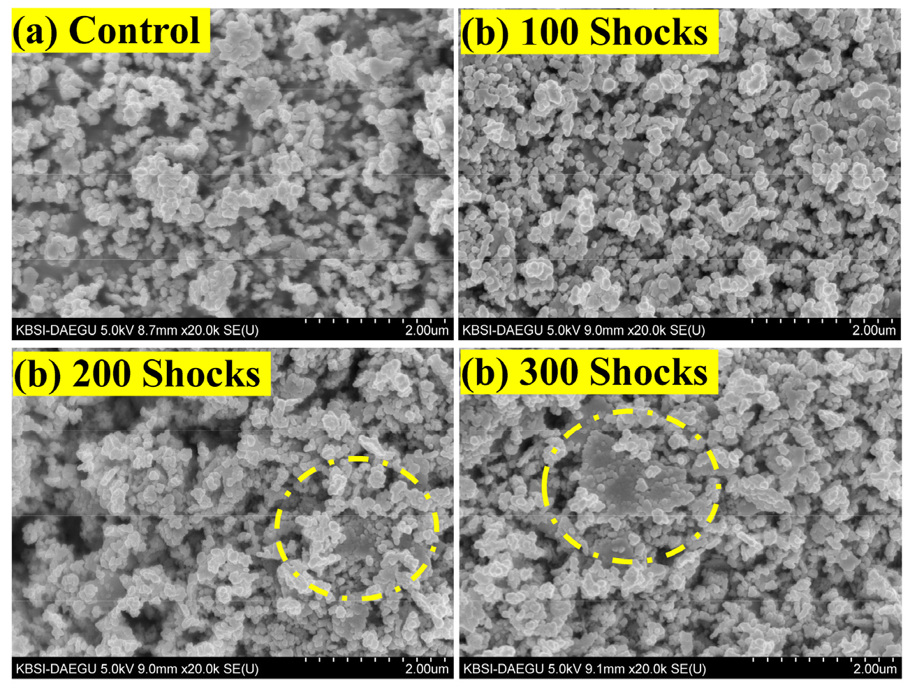
The morphological analysis confirmed that the synthesized ZnO nanorods exhibit excellent stability under shock wave-loaded conditions, with no notable defects or shape alterations [30]. This remarkable stability of the ZnO NRs can be attributed to the strong affinity between the zinc and oxygen atoms, as well as their shorter bond length. In contrast, previous studies have reported that titanium dioxide nanoparticles (TiO2 NPs) are less stable against shock waves, undergoing phase transformations under such conditions, and ZnO (ZnO NPs) is stable for shock-loaded conditions. Consequently, ZnO NRs are proposed as suitable candidates for aerospace and military applications due to their outstanding shock wave-resistant properties.
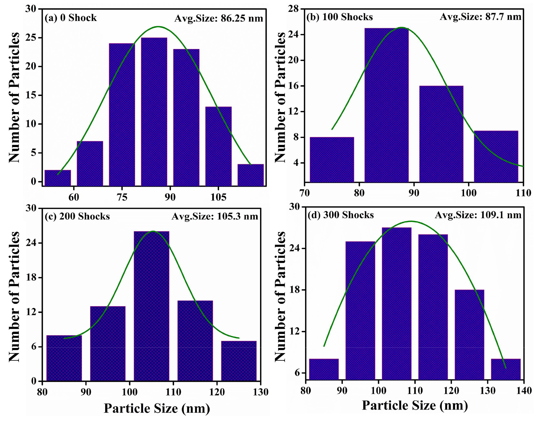
From the morphology, we calculated the particle size for the control and shock-loaded samples as shown in Fig. 6(a)-(d). FESEM images enable a detailed examination of a material’s morphological characteristics, including particle size, shape, and surface features. The distribution, clustering, or aggregation of particles, which can impact the material’s behavior, are also visible. In some cases, the images reveal information about the internal structure, such as crystallinity and grain boundaries which plays a crucial role in their properties. In the present experiment, for more accurate and reliability, we have used ‘ImageJ’ software for particle size calculations. As the number of shock pulses increases, the average particle size correspondingly increases, reaching 109.1 for 300 shocks compared to 86.25 for the control sample.
As discussed, shock wave propagation can cause smaller particles to fuse, leading to larger agglomerations. This fusion effect increases the effective particle size as individual particles cluster into larger sizes [31]. Additionally, Fig. 6(c), (d) indicates a sheet-like structure, which is attributed to the high strain rates from shock waves. This can lead to dynamic recrystallization, where smaller grains merge or grow under the influence of heat and pressure. This growth of grains contributes to an apparent increase in particle size [32,33].
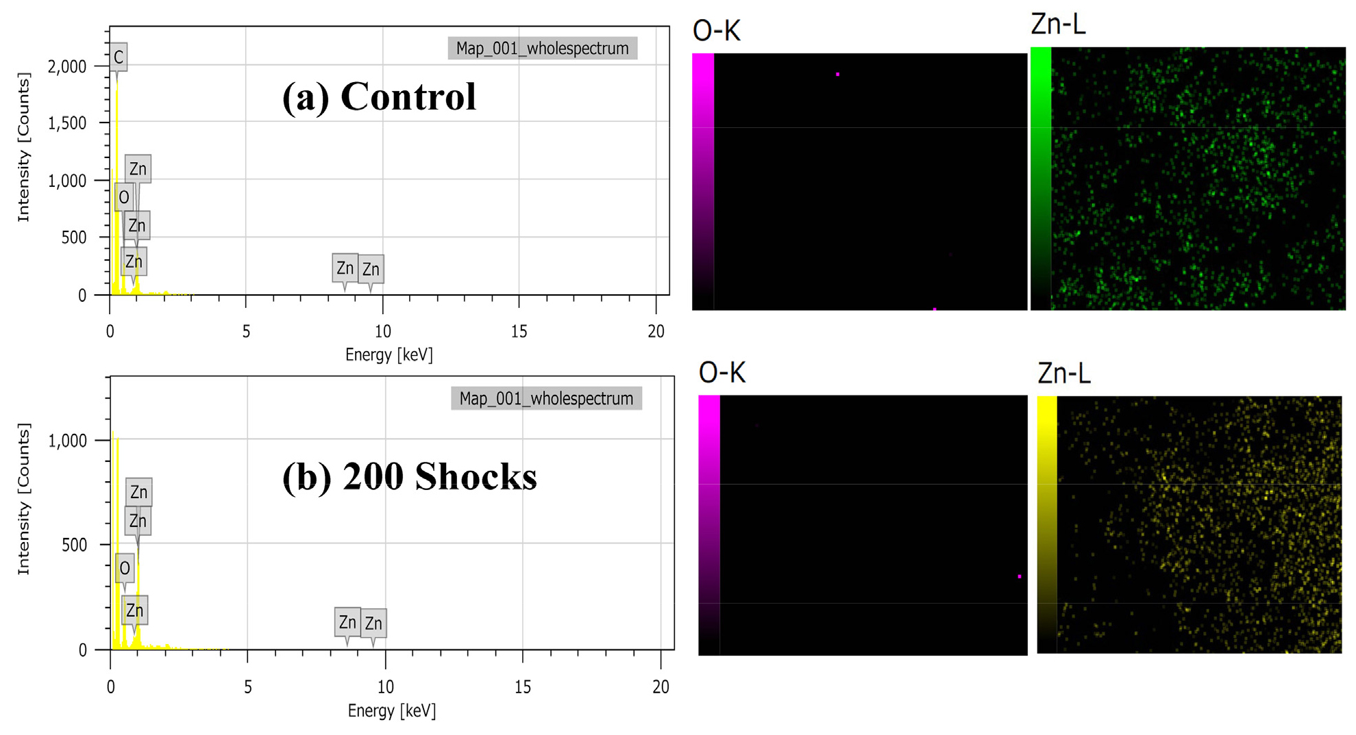
For the elemental confirmation, we performed energy-dispersive x-ray analysis (EDAX) analysis and elemental mapping on the control and 200 shock samples as shown in Fig. 7. The results reveal that the ZnO material contains only the expected zinc and oxygen elements, with no impurities observed for both samples. This analysis confirms the purity and composition of the ZnO material used in the study.
4. Conclusions
This study’s findings highlight the remarkable stability of ZnO nanoparticles when subjected to shock-loaded conditions. The retention of their hexagonal crystalline structure and the observed agglomeration and growth in particle size demonstrate ZnOs ability to withstand shock waves without structural degradation. This points to its potential utility in aerospace and military applications, where materials must contend with extreme conditions such as 100, 200, and 300 shocks. The insights gained from powder diffraction and morphological analysis provide a foundational understanding of ZnOs performance, paving the way for further exploration and application in extreme environments. Overall, ZnO nanoparticles present a promising option for enhancing the resilience of critical aerospace technologies.
